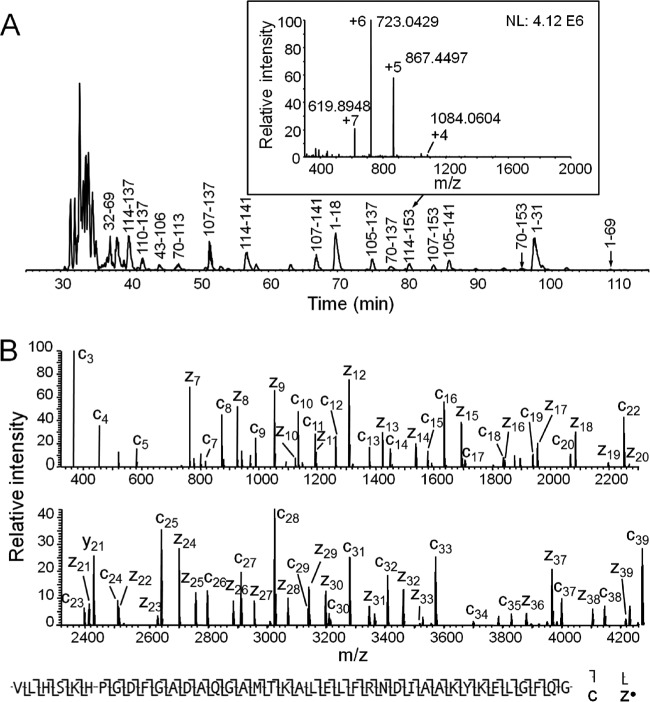
Fig. 2.
A, Base peak chromatogram of apomyoglobin digest generated by 0.77 s time-controlled digestion, with the 3–9 kDa base peak peptides labeled using apomyoglobin sequence number. Inset is the MS1 spectrum at the elution time corresponding to peptide 114–153, and ions corresponding to peptide 114–153 are labeled with different charge states. B, The spectrum of apomyoglobin peptide 114–153 after converting the original ETD MS/MS spectrum to +1 ions by Xcalibur Xtract (some fragment ions are lost after Xtract convertion). Under the spectrum is the sequence coverage by c and z· ions assigned by ProSite PC 3.0 using the original MS/MS data (manually verified).

