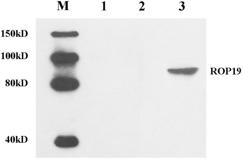
Figure 3.
In vitro expression analysis of the constructs in HEK 293-T cells by Western blot. Protein marker (lane M), untransfected cells (lane 1), cells transfected with pEGFP-C1 (lane 2), and cells transfected with pROP19 (lane 3). The marker contains 150 kD, 100 kD, 80 kD, and 40 kD. The detected band is located between 80 kD and 100 kD.

