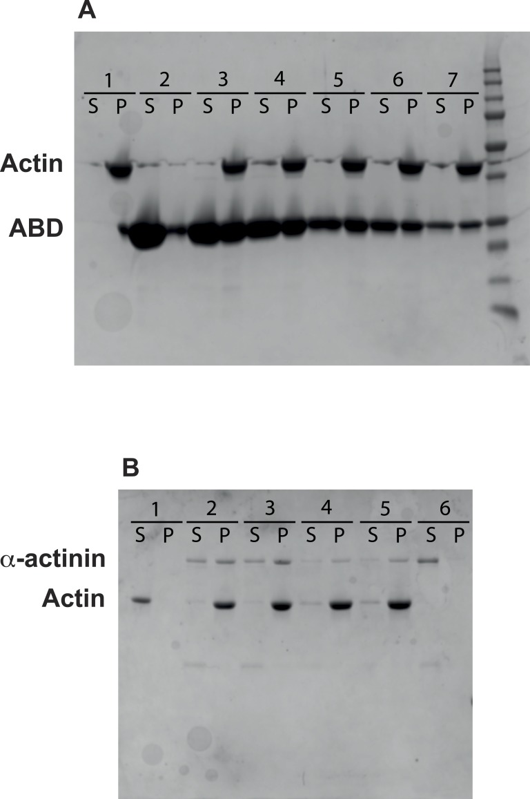
Figure 6. Actin binding and bundling.
(A) Human platelet non-muscle actin was mixed with varying concentrations of ABD, incubated at room temperature for 60 min and centrifuged for 60 min@ 90,000 rpm. Supernatants and pelleted proteins were analysed by SDS-PAGE. Lane 1: 12 µM actin; lane 2: 251 µM ABD; lane 3: 12 µM actin and 251 µM ABD; lane 4: 12 µM actin and 126 µM ABD; lane 5: 12 µM actin and 63 µM ABD; lane 6: 12 µM actin and 31 µM ABD; lane 7: 12 µM actin and 16 µM ABD. (B) Actin was mixed with full length α-actinin, incubated as before and centrifuged for 11 min@ 13,000 rpm. Supernatants and pelleted proteins were analysed by SDS-PAGE. Lane 1: 5 µM actin; lanes 2 and 3: 5 µM actin and 3.7 µM α-actinin; lanes 4 and 5: 5 µM actin and 1.2 µM α-actinin; lane 6: 3.7 µM α-actinin.

