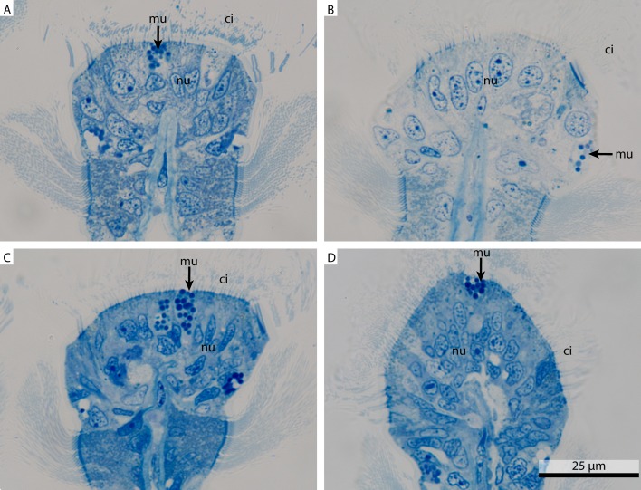
Figure 1. Light micrograph comparing four differently fixed tissue pieces of Mytilus edulis.
Overview of four differently fixed Mytilus edulisgills. All images show transverse sections through gill filaments focusing on the ciliated frontal surface. (A) shows a FSW fixed sample, (B) a marCaco fixed sample, (C) a marPHEM fixed sample and (D) a marPBS fixed sample. ci, cilia; mu, mucus granule; nu, nucleus.

