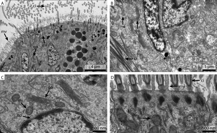
Figure 4. Ultrastructural details of marPHEM fixed gill cell of Mytilus edulis.
(A) shows an overview of the cells of the ciliated frontal surface, (B) is a higher magnification of the same, (C) shows the nucleus in higher magnification and (D) shows details of the cell surface. go, Golgi apparatus; nu, nucleus; ci, cilia; mu, mucus granule; mv, microvilli; np, nuclear pore; cr, ciliary root; rer, rough ER; mi, mitochondria.

