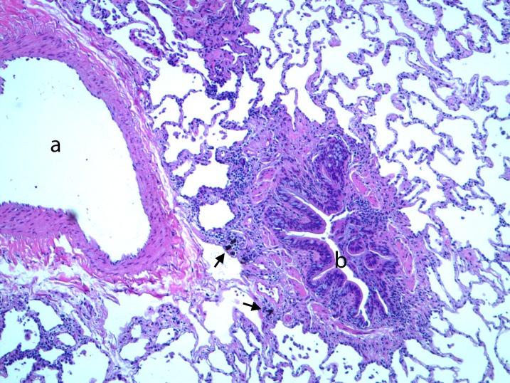
Figure 1.
Representative hematoxylin and eosin–stained cryobiopsy diagnostic of constrictive bronchiolitis. The bronchiolar wall is abnormally thickened by a combination of smooth muscle hyperplasia and collagen deposition. There is an adventitial chronic inflammatory infiltrate and deposition of black pigment in the bronchovascular notch (arrows). The lumen (b) contains prominent infoldings and is smaller in diameter than the accompanying artery (a). There is prominent goblet cell metaplasia (×100).

