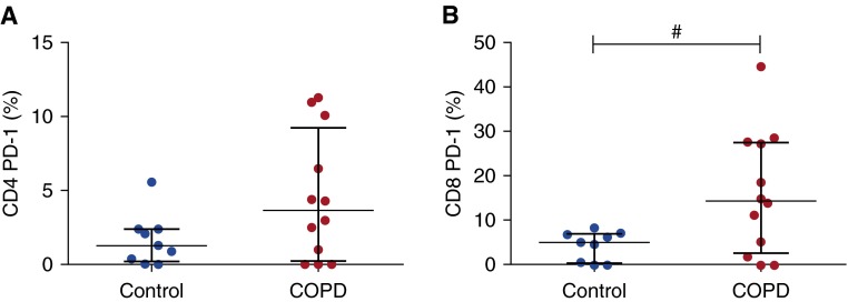
Figure 2.
Intrinsic programmed cell death protein (PD)-1 expression by CD4 and CD8 T cells in controls and chronic obstructive pulmonary disease (COPD). After explanted lung tissue was rested overnight, tissue was digested with collagenase and cells were analyzed by flow cytometry. T cells are gated on the live CD45+CD3+ population. Proportions of (A) CD4+ and (B) CD8+ T cells expressing surface PD-1 are shown (control, n = 9; COPD, n = 12). Median and interquartile range are shown. Data were analyzed using the Mann-Whitney U test (#P < 0.05). Note that the y-axis limit is changed from 15% in A to 50% in B.

