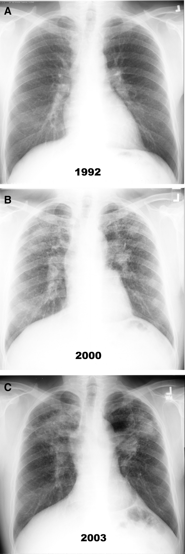
Figure 2.
Posteroanterior radiographs of a coal miner with rapidly progressive pneumoconiosis who underwent a bilateral lung transplant at age 60 years (see pathology in Figure 3). He had 35 years of coal-mining experience, of which 28 years were at the face of the mine, and no history of smoking. (A) Chest radiograph obtained after 24 years of coal mine employment showing category 3 simple pneumoconiosis with q- and r-type opacities. (B and C) Chest radiographs obtained after 32 and 35 years of coal-mining employment, respectively. Multiple, large, mass-like lesions and nodules in the bilateral upper lung fields, which coalesce further and enlarge as time progresses, are shown.

