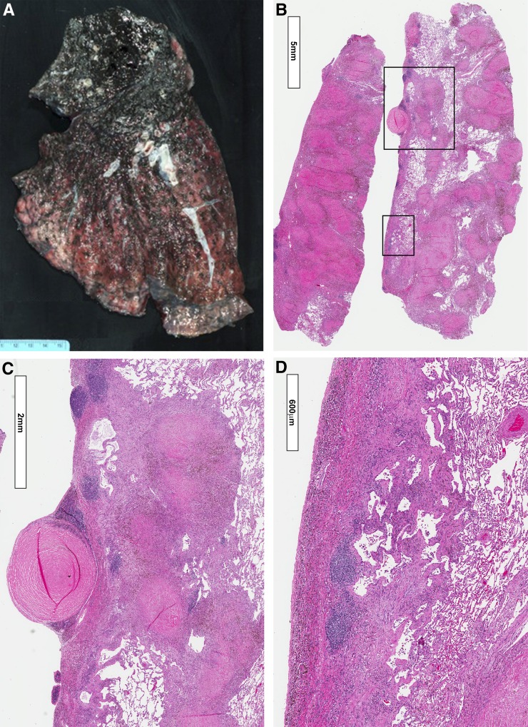
Figure 3.
Explanted left lung from the miner whose radiographs are shown in Figure 2. (A) Photograph of the lung explant. The upper lobe is completely replaced by progressive massive fibrosis (PMF). Pale nodular areas can be seen within the PMF, indicative of silicosis. The apical segment of the lower lobe is also involved with PMF. Elsewhere the lung shows nodular lesions of pneumoconiosis. (B) Low-magnification view of section from area of PMF (left) and area involved with simple coal workers’ pneumoconiosis (right). Both areas show predominantly silicotic lesions. (C) Close-up of upper boxed area shown in B. A silicotic nodule can be seen on the pleural surface (pleural pearl), together with subpleural, semiconfluent, silicotic nodules. There is a marked lymphoid reaction in the pleura. (D) Close-up of lower boxed area in B showing pleural fibrosis with shallow underlying interstitial fibrosis as well as lymphoid aggregates.

