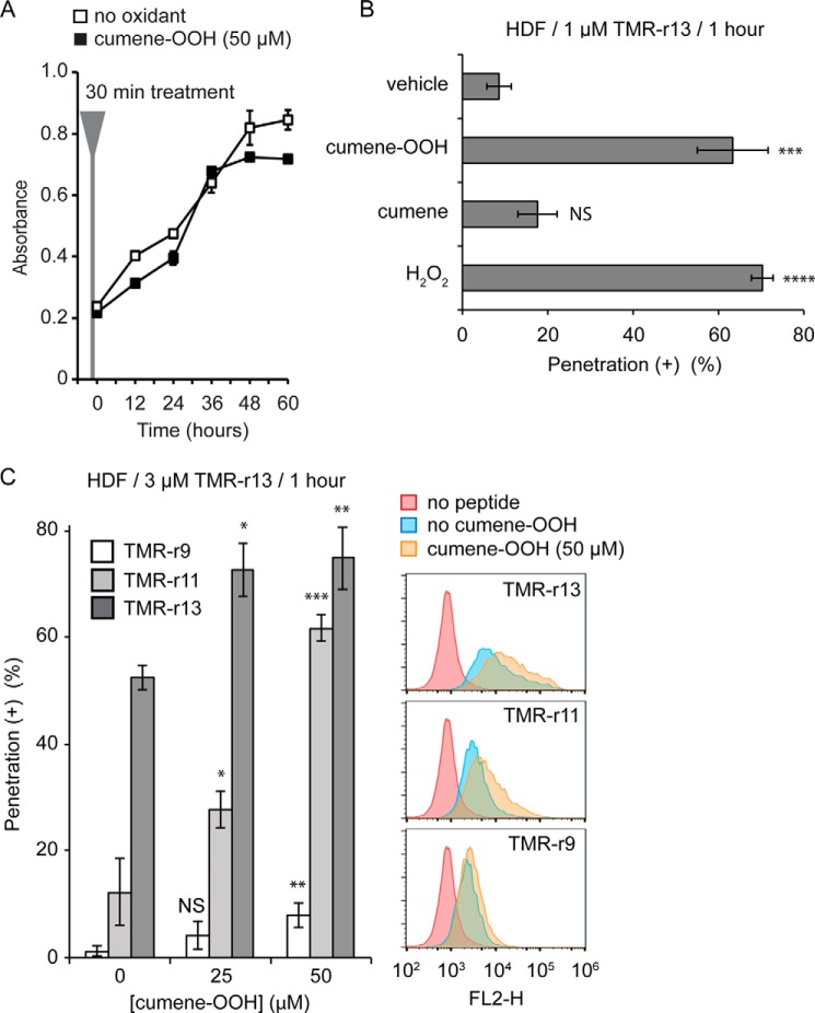
FIGURE 4.
Oxidants increase the cytosolic penetration of TMR-r13. A, HDF cells treated with cumene-OOH (50 μm, 30 min) display a proliferation rate similar to untreated cells. B, cell penetration activity of TMR-r13 in HDF increases significantly in the presence of oxidants. HDF cells were pretreated with cumene-OOH, cumene, or H2O2 for 30 min and incubated with 1 μm TMR-r13 for 1 h. *** represents p ≤ 0.001; **** represents p ≤ 0.0001, and NS represents p > 0.05 compared with vehicle (PBS). C, cumene-OOH (0, 25, or 50 μm, 30 min) increases the activity of TMR-r13, TMR-r11, and TMR-r9 (3 μm, 1 h) in a dose-dependent manner in HDF cells. Cytosolic penetration was quantified by fluorescence microscopy (histogram) and flow cytometry. NS represents p > 0.05; * represents p ≤ 0.05; ** represents p ≤ 0.01, and *** represents p ≤ 0.001 compared with cells not treated with cumene-OOH. FL2-H is signal intensity of sample in the FL2 channel (Ex = 488 nm/Em = 585 ± 40 nm, used for detection of TMR-r13). The data in A–C represent the mean of triplicate experiments and the corresponding standard deviations.

