FIGURE 5.
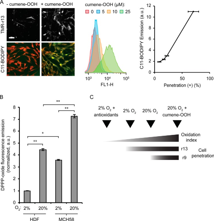
Membrane oxidation and cytosolic penetration are positively correlated. A, HDF cells were treated with cumene-OOH at various concentrations for 30 min. The cytosolic penetration efficiency of TMR-r13 (1 μm, 1 h) was measured. Images of TMR-r13 delivery are monochromes of TMR fluorescence emission (RFP filter). In parallel, membrane oxidation after cumene-OOH treatment was measured by flow cytometry using the green fluorescence of the lipophilic oxidation reporter C11-BODIPY581/591. FL1-H corresponds to the signal intensity of sample in the FL1 channel (Ex = 488 nm/Em = 533 ± 30 nm, used for detection of the green fluorescence of C11-BODIPY581/591). The correlation between the geometric mean of the C11-BODIPY581/591 emission and the cytosolic penetration efficiency is reported. Scale bar, 20 μm. B, comparison of the levels of lipid peroxides in the membrane of HDF and MCH58 cells using the lipophilic DPPP probe. The fluorescence of oxidized DPPP present in cellular membranes of cells grown at 2% or 20% oxygen is reported as normalized to the total number of cells per sample and to the least oxidized sample (2%, HDF). * represents p ≤ 0.05, and ** represents p ≤ 0.01. C, scheme illustrating the relationship between the cellular penetration of TMR-r13 and TMR-r9 with membrane oxidation index.

