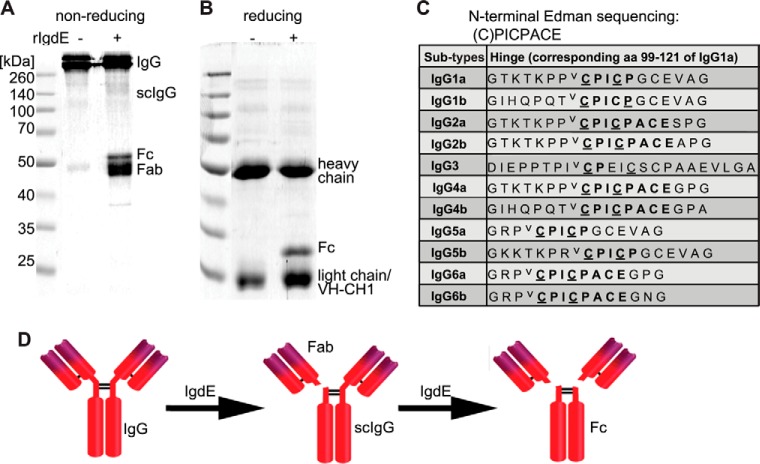
FIGURE 4.
IgdE cleaves the heavy chain of porcine IgG in the hinge region. The reaction was analyzed by non-reducing (A) and reducing (B) Coomassie Blue-stained SDS-PAGE. 3.3 μm IgG were incubated with (+) or without (−) 10 nm purified rIgdE for 16 h at 37 °C. C, hinge region sequences of all porcine IgG subtypes. Cysteine residues believed to be involved in S-S covalent bonds (underlined) and the potential cleavage site (⋁) are marked in the table. D, the observed cleavage pattern and cleavage site proposes a model where one IgG heavy chain is first hydrolyzed by IgdE resulting in one free Fab fragment (Fab) and single cleaved IgG (scIgG) and in a second step the other heavy chain is hydrolyzed resulting in one Fc fragment (Fc) and two Fab fragments.

