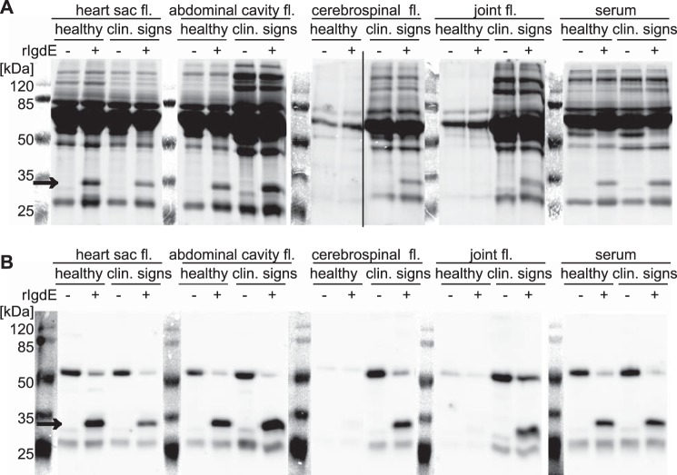
FIGURE 6.
IgdE degrades endogenous IgG in all tested body fluids of healthy pigs and pigs with respective lesions (size indicated with arrows). No other degradation products could be observed. Diluted body fluid samples were incubated with (+) or without (−) 10 nm purified rIgdE for 16 h at 37 °C. The reactions were analyzed by SDS-PAGE (A) and anti-IgG Western blots (B) under reducing conditions. Lanes showing the protein size standard are a photographic image of the membrane before detection of the chemiluminescence signal. Images of different gels have been assembled into one figure.

