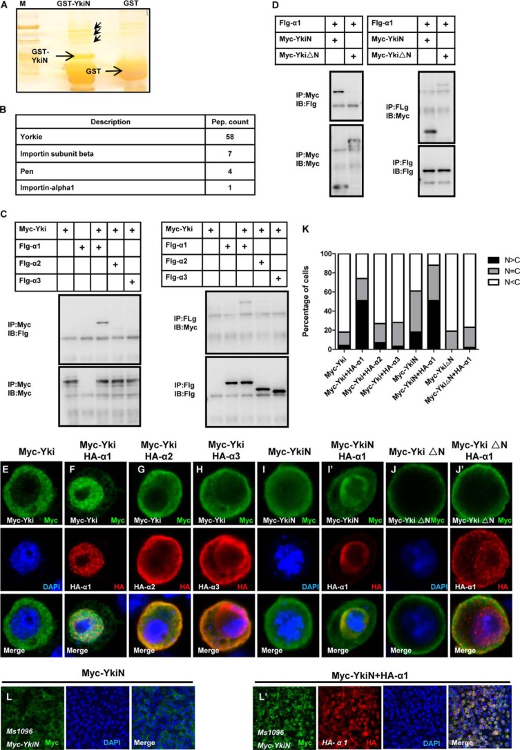
FIGURE 2.
Importin α1 binds to the N terminus of Yki and mediates the nuclear localization of Yki. A, silver staining of the sample for MS. GST and GST-YkiN protein were purified and coupled with GST beads, then pull-down using the S2 cell lysate. The specific bands are shown with red arrowhead. B, results for MS. MS analysis have identified Importin α1, pen (Importin α2), Importin β. C, Importin α1 interacts with Yki in vitro. S2 cells were transfected with the indicated constructs. The cell lysates were immunoprecipitated and subjected to Western blot analysis. D, Importin α1 only interacts with YkiN. S2 cells expressing Myc-YkiN, Myc-YkiΔN, and Flg-Importin α1 were immunoprecipitated and probed with the indicated antibodies. E–H, S2 cells expressing Myc-Yki (E) or Myc-Yki with HA-Importin α1 (F), HA-Importin α2 (G), HA-Importin α3 (H) were immunostained with anti-Myc (green) or anti-HA (red) antibodies. I–J′, S2 cells expressing Myc-YkiN (I–I′), Myc-YkiΔN (J–J′) with or without HA-Importin α1 were immunostained with anti-Myc (green) or anti-HA (red) antibodies. Nuclei are marked by DAPI (blue). K, cells with different nucleocytoplasmic distributions of Myc-tagged variants were counted. A total of 100 cells were counted for each case. The y axis indicates the percentage of cells in each category. L–L′, Drosophila imaginal wing discs expressing Myc-YkiN (L) or Myc-YkiN plus HA-Importin α1 (L′) by MS1096 were dissected and stained with anti-Yki (green) or anti-HA (red) antibodies.

