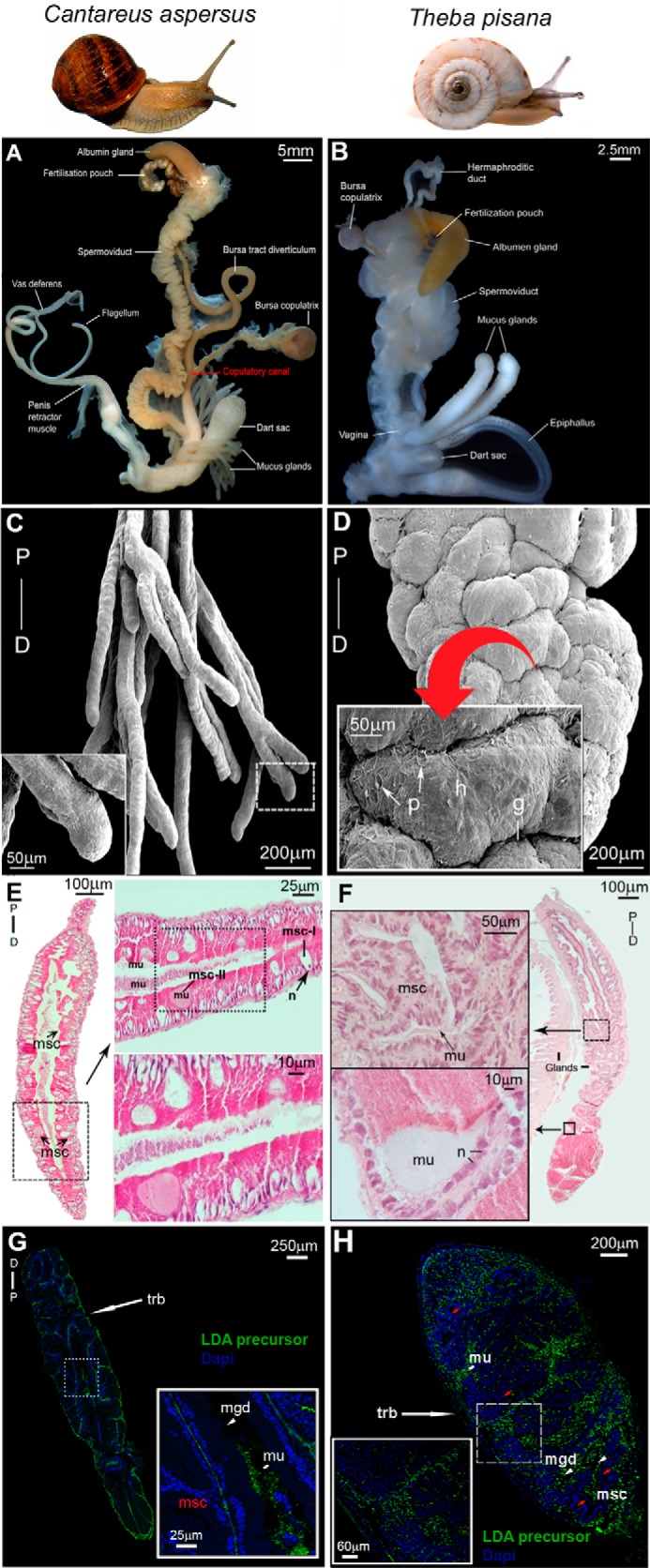
FIGURE 6.

Localization of LDA in mucous glands of C. aspersum and T. pisana. A and B, C. aspersum (A) and T. pisana (B) reproductive systems, including the bursa copulatrix involved in degrading sperm, the copulatory canal (observed in C. aspersum), which is the major site that directs the closure of the aperture that allows for sperm storage and/or sperm degradation at the BC, and paired mucous glands with associated dart sac. C and D, scanning electron micrographs of one branch of the mucous gland pair of C. aspersum (C) and T. pisana (D). P, proximal; D, distal. Higher power micrographs are shown (inset). In T. pisana, convoluted ridges/hillocks (h) are separated by serrated grooves (g) and pits (p) of ∼50 μm. E, hematoxylin and eosin histology of the C. aspersum mucous gland showing msc. Higher magnifications are shown as horizontal insets that demonstrate mucus-deficient cells (msc-I) with a distinct nucleus (n), and mucus-bearing cells (msc-II) rich with mucus (mu). F, hematoxylin and eosin histology of the T. pisana mucous gland showing a similar anatomical arrangement of msc with stores of mucus. Higher power micrographs are shown as insets. G and H, immunolocalization of LDA precursor in C. aspersum and T. pisana mucous glands. Both show immunoreactivity at the region of the outer trabeculae (trb) and in mucous gland ducts (mgd).
