
FIGURE 2.
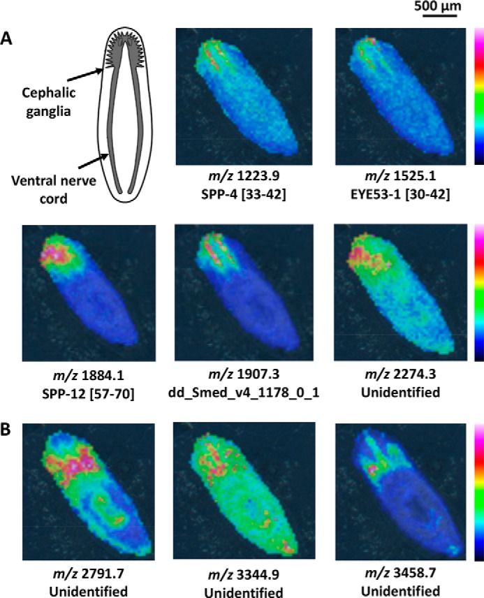
MALDI MSI ion images for intact planarians. A, many peptides are localized to the cephalic ganglia or toward the anterior end of the planarian. Peptides derived from different SPP are labeled with their sequence portion within the prohormone. Peptides not derived from known prohormones are labeled with their FASTA annotation as entered in the planarian transcriptome. Unidentified ions were detected via MALDI MSI but were not characterized in the follow-up HPLC-ESI MS/MS peptidomic analysis. The diagram of the planarian nervous system shown in the upper left was modified from a previous study (71). B, peptides that are localized in the mesenchyme around the cephalic ganglia are also observed. The anterior of each animal is positioned toward the top left. Signal intensity is color coded, with intensity scales provided. Intensity increases from blue to red.