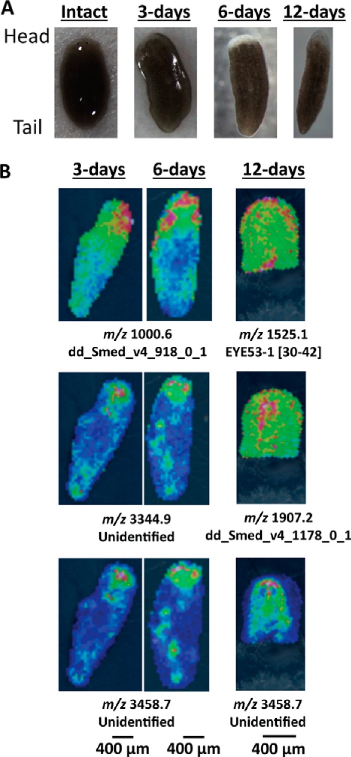
FIGURE 3.

Optical and ion images for regenerating planarians at 3, 6, and 12 days after amputating the cephalic ganglia. A, optical images of intact and regenerating planarians. B, several ions are localized toward the blastema in 3-day and 6-day regenerating planarians. The cephalic ganglia are visible after 12 days of regeneration. In 12-day regenerates, only the anterior half of the planarian is imaged. Peptides derived from known prohormones, such as EYE53-1, are labeled with their sequence portion within the prohormone. Peptides that are not derived from known prohormones are labeled with their FASTA annotation as entered in the planarian transcriptome. Unidentified ions were detected via MALDI MSI but were not characterized in the follow-up HPLC-ESI MS/MS peptidomic analysis. Anterior is to the top for each panel. Signal intensity is color coded, with intensity scales provided. Intensity increases from blue to red.
