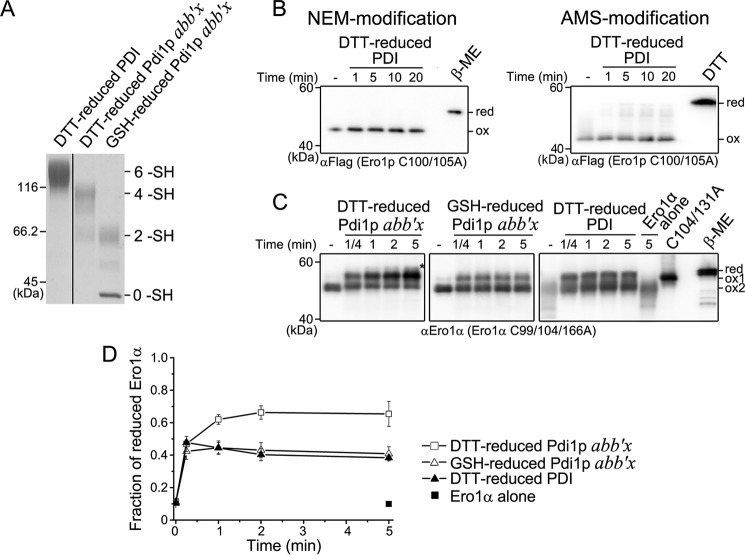
FIGURE 7.
Complementary activation of Ero1p and Ero1α by PDI and Pdi1p. A, reduced PDI and Pdi1p were prepared and monitored as described in the legend to Fig. 2A. The number of free thiols (-SH) in PDI and Pdi1p were labeled on the right margin. B, the reduction of the long-range (left) and short-range (right) regulatory disulfides in Ero1p C100A/C105A-FLAG by reduced PDI was monitored as described in the legend to Fig. 2, B and D, respectively. C, the activation of catalytically inactive Ero1α variant C99A/C104A/C166A retaining both long-range disulfides of Cys85-Cys391 and Cys94-Cys131 (18). Oxidized Ero1α C99A/C104A/C166A (ox2) at 0.5 μm was incubated in the absence or presence of 10 μm reduced PDI or Pdi1p as indicated. Aliquots were taken at the indicated times, and the redox states of NEM-blocked Ero1α were analyzed under non-reducing conditions by Western blotting using αEro1α rabbit serum. Ero1α C104A/C131A was loaded as an indicator for the reduction of Cys94-Cys131 disulfide (ox1). Fully reduced Ero1α C99A/C104A/C166A was prepared with excess β-mercaptoethanol (β-ME) (red). The asterisk indicated Ero1α species with both Cys85-Cys391 and Cys94-Cys131 disulfides reduced (18). D, the fraction of activated Ero1α (ox1 and red) in C was quantified by densitometry and plotted against time (mean ± S.D., n = 3 independent experiments).

