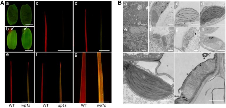
Figure 2.
Chlorophyll autofluorescence and TEM observations of wild type and wp1s. A, Observation of autofluorescence in wild-type and wp1s seedlings. Seeds of wild type and wp1 before (a) and 2 d after imbibition (b). c-g, Leaf tips of wild type and wp1s at P4-2 stage (c), P4-4 stage (d), P4-8 stage (e), P4-10 stage (f), and mature fourth leaf stage (g). Scale bars = 3 mm. B, Transmission electron micrographs of plastids in wild-type and wp1s seedlings. a, B, c, D, I, Wild type. e, F, g, H, j, wp1s. Tissues were collected from the seedlings at the following developmental stages: embryo (a, e), P4-2 (b, f), P4-4 (c, g), P4-8 (d, h), and P4-10 (i, j). Scale bars = 2 μm (a, e), 0.5 μm (b, f), and 1 μm (c, D, g, F, i, j).

