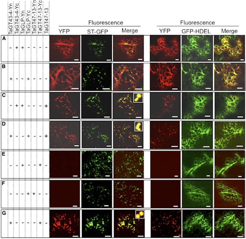
Figure 5.
TaGT43-4, TaGT47-13, and TaGLP assemble in the ER before export to the trans-Golgi. Protein-protein interactions were visualized via BiFC (split-YFP). Coinfiltrated constructs are indicated by the “+” at the left of the figure. ER marker (GFP-HDEL) and trans-Golgi marker (ST-GFP) were included to show the localization of the reconstituted YFP. GFP and YFP fluorescence are shown in green and red, respectively, and their colocalization (merge) appears in yellow. The insets in merge pictures in C, D, and G show the overlap between YFP and ST-GFP. Bars = 10 μm

