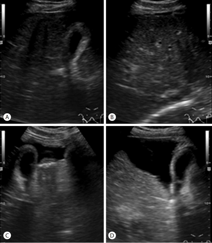
Figure 1.

Abdominal ultrasonographic findings. Intercostal and transverse sonograms (A, B) show coarse parenchymal echogenicity, surface nodularity, and a moderate amount of ascites in the perihepatic space. Subcostal oblique sonograms (C, D) show a large amount of ascites in the widened interlobar fissure, which is considered a typical finding of liver cirrhosis.
