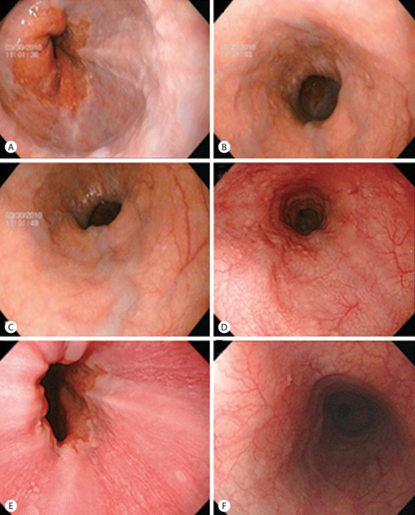
Figure 2.

Esophagogastroduodenoscopic findings. Straight to slightly enlarged (A, B) and tortuous varices (C) were observed on the lower esophagus. The esophageal varices had decreased to minimal varices after 2 years of entecavir therapy (D), and had completely disappeared after 4 years of entecavir therapy (E, F).
