
Fig. 2. Ex vivo and in vivo investigation of the mechanism of drug delivery enhancement with ultrasound.
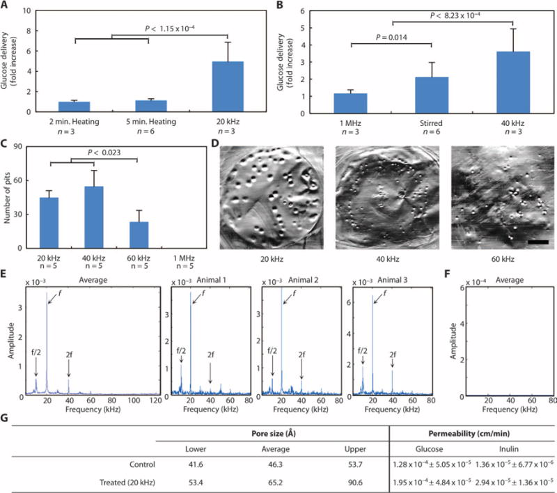
(A) Relative enhancement in glucose delivery to small intestine as a result of 40°C heating for 2 or 5 min or 20-kHz ultrasound at 7.5 W/cm2 at a duty cycle of 50% for 2 min total. (B) Relative enhancement in glucose delivery to small intestine as a result of treatment with 1-MHz ultrasound set to 2 W/cm2 (5.22 W actual) for 3.4 min, stirring of the donor chamber, or 40-kHz ultrasound set to an intensity of 13.4 W/cm2. (C) Number of pits generated in aluminum foil after treatment with 20-, 40-, 60-kHz, or 1-MHz ultrasound for 2 s at the highest intensity considered for each frequency. In (A) to (C), data are averages ± 1 SD; P values were determined by one-way analysis of variance (ANOVA) with multiple comparisons. (D) Representative images of pitted aluminum foil samples treated with either 20-, 40-, or 60-kHz ultrasound. Scale bar, 3 mm. (E and F) Averaged Gaussian filtered amplitude spectrum of six fast Fourier transforms (FFTs) taken on in situ acoustic data collected externally in vivo in pigs during f = 20 kHz UMGID along with a representative amplitude spectrum for each repeat (E) and a control when ultrasound was not on (F). (G) Estimation of pore size radii created in porcine small intestine tissue and permeability (averages ± 1 SD) of glucose and inulin as a result of ultrasound exposure using the aqueous porous pathway model.