
Fig. 4. Effect of rectal ultrasound on blood markers, histology, fecal score, and cytokine expression.
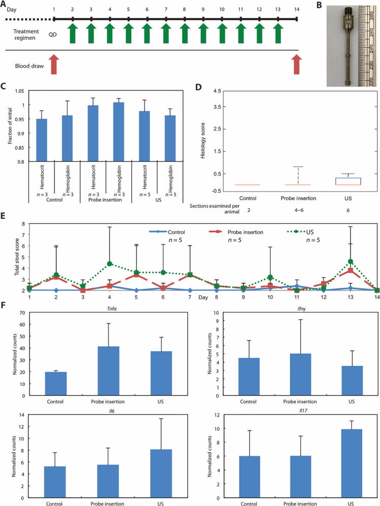
(A) Daily ultrasound treatment schedule in healthy pigs in the absence of colitis. (B) The custom-designed ultrasound probe tip with a shaft diameter of 2 mm. The two bumps shown have a diameter of 3 mm and enhance radial ultrasound emission. (C) Hematocrit and hemoglobin normalized to day 1 for healthy animals (control), healthy animals receiving daily probe insertion (probe insertion), and healthy animals receiving daily 40-kHz ultrasound treatment (US). Although five animals were used in each group, some blood samples from days 1 and 14 clotted, resulting in fewer than five values for some groups. (D) Histology scores of tissue sections at day 14. The median, 25th, and 75th percentiles are shown. The whiskers indicate the most extreme data points. (E) Total fecal score for all three groups over the 14-day period. (F) Cytokine mRNA levels in colonic tissue samples (n = 4 biological repeats for all groups). Counts were assessed using the Mouse Inflammatory Panel (NanoString Technologies). Data are normalized across samples using internal positive spike-in controls. Data in (C), (E), and (F) are averages ± 1 SD. Sample sizes indicated are biological replicates.