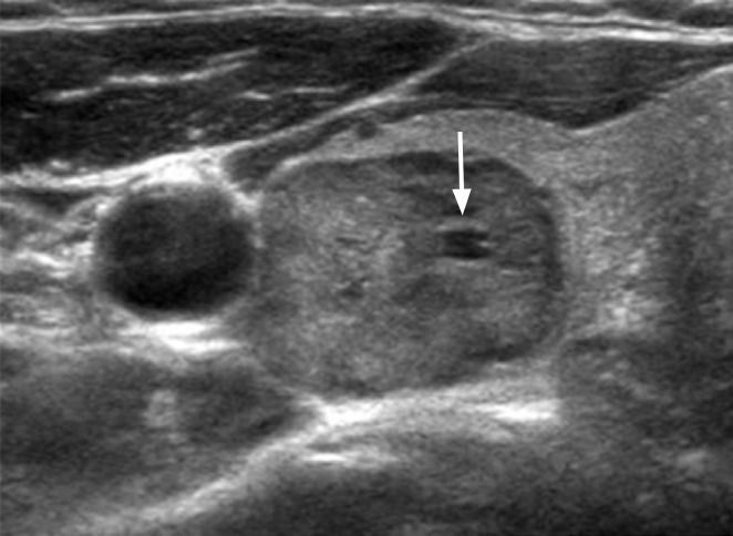
Fig. 2. A hypoechoic nodule with minimal cystic changes in a 50-year-old woman.

The mildly hypoechoic nodule (30 mm) has a smooth margin, ovoid shape, parallel orientation, and focal minimal cystic change (arrow). The final diagnosis was benign follicular nodule.
