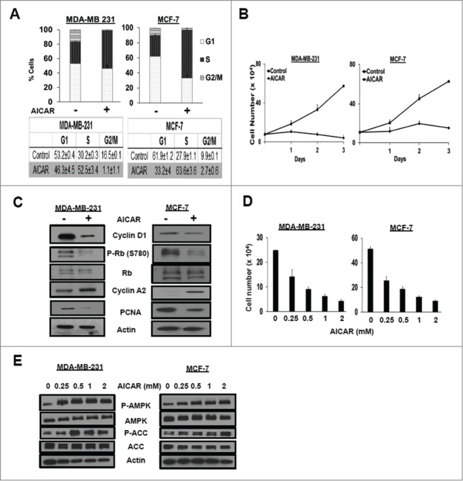
Figure 1.

AICAR treatment causes S-phase cell cycle arrest. (A) MDA-MB-231 and MCF-7 cells were plated at 30% confluence in 10-cm plates in DMEM containing 10% serum. After 24 hr, the cells were treated with AICAR (0.5 mM) for 48 hr. After 48 h, the cells were harvested, fixed, stained with propidium iodide, and analyzed for cell cycle distribution by measuring DNA content/cell as described in Methods. The error bars represent the standard error of the mean for experiments repeated 3 times. (B) Cells were plated at 20% confluence in 6-well plates in complete media containing 10% serum. After 24 hr, AICAR (0.5mM) was added. Cells were harvested at indicated time points, stained using crystal violet, and quantified by light microscopy as described in Methods. Error bars represent the standard error for an experiment repeated 3 times. (C) Cells were plated at 30% confluence in 10-cm plates in complete media containing 10% serum for 24 h at which time they were treated with AICAR (0.5 mM) for 48 hr. The cells were subsequently harvested and cell lysates were collected. The indicated protein levels were determined by Western blot analysis. The data shown are representative of experiments repeated at least 2 times. (D) Cells were seeded as in B and treated with various concentrations of AICAR (0.25–2mM) for 48 hr, at which time the cells were harvested, stained using crystal violet, and quantified by light microscopy as described in Experimental Procedures. Error bars represent the standard error for an experiment repeated 3 times. (E) Cells were seeded as in (C)and treated with various concentrations of AICAR (0.25–2mM) for 24 hr. Cells were harvested and the levels of phospho-AMPK, AMPK, phospho-acetyl-CoA carboxylase (P-ACC), ACC, and actin were determined by Western blot analysis. The data shown are representative of experiments repeated at least 2 times.
