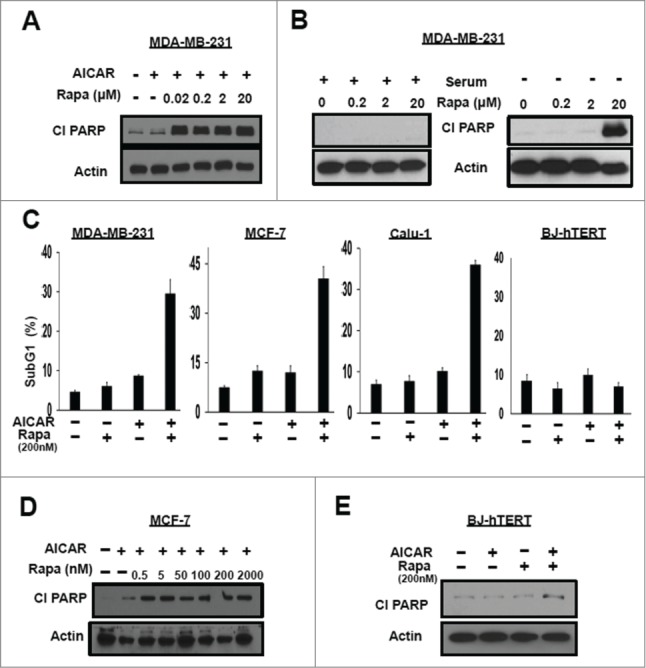
Figure 2.

AICAR treatment reduces the concentration of rapamycin to induce apoptosis. (A) MDA-MB-231 cells were plated at 60% confluence in 60mm plates in DMEM containing 10% serum. Twenty-four hr later the cells were treated with AICAR (2mM) and/or different doses of rapamycin as indicated for 24 hr. The cells were then harvested and levels of cleaved PARP (Cl PARP) and actin were determined by Western blot analysis. (B) MDA-MB-231 cells were plated as in A. Twenty-four hr later of plating, the cells were shifted to complete medium or medium lacking serum and treated with rapamycin at different doses for 24 hr. The cells were then harvested and indicated protein levels were determined as in A. (C) MDA-MB-231, MCF-7, Calu-1 and BJ-hTERT cells were plated at 40% confluence and treated with AICAR (0.5 mM) and/or rapamycin (200 nM) for 48 hr, after which these were collected and subjected to flow cytometric analysis. Total subgenomic DNA is plotted as indicated. Error bars represent SD values for at least 2 independent experiments. (D) MCF-7 cells were plated in A. The cells were treated with AICAR (2 mM) and/or varying doses of rapamycin as indicated for 24 hr. The cells were then harvested and indicated protein levels were determined as in A. (E) BJ-hTERT cells were plated in A. The cells were treated with AICAR (2 mM) and/or rapamycin (200 nM) for 24 hr. The cells were then harvested and indicated protein levels were determined as in A. The data shown are representative of experiments repeated at least 2 times.
