
Figure 4.
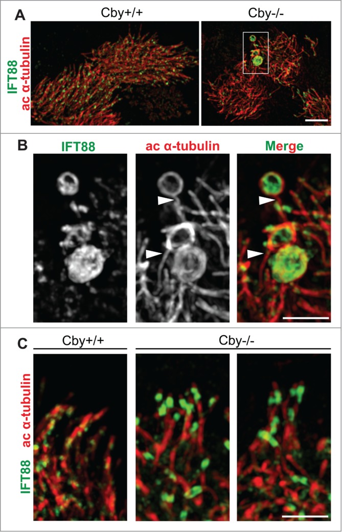
IFT88 accumulations are contained within the bulge structures at the dilated ciliary tips of Cby−/− tracheal ciliated cells. (A) Super-resolution 3D-SIM images of ALId21 Cby+/+ and Cby−/− MTECs colabeled for IFT88 (green) and ac α-tubulin (red). Scale bar: 5 µm. (B) Magnified images of the boxed area in (A) clearly demonstrate that IFT88 accumulations occurred within dilated ac α-tubulin-positive enclosures at the ciliary tip. Single channel images for IFT88 and ac α-tubulin staining are shown in grayscale. Ciliary axonemes, which harbor IFT88 accumulations, are indicated by arrowheads. Scale bar: 1 µm. (C) 3D-SIM images of cilia in Cby+/+ and Cby−/− MTECs at ALId21 colabeled for IFT88 (green) and ac α-tubulin (red). Moderate IFT88 accumulations at the ciliary tip were frequently found in Cby−/− ciliated cells. Note that IFT88 accumulations, large or small, were not seen in Cby+/+ cilia. Scale bar: 1 µm.