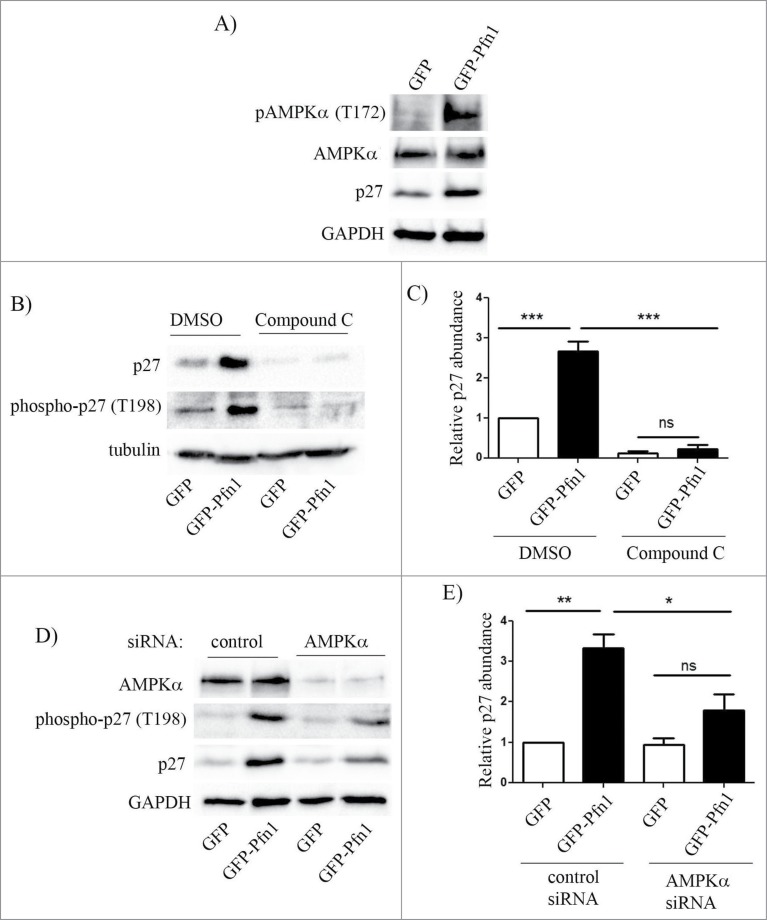
Figure 4.
Pfn1 overexpression upregulates p27 in MDA-231 cells through AMPK activation. (A) Immunoblots of total extracts show the relative levels of T172-phosphorylated- AMPK, total AMPK and p27 between GFP- and GFP-Pfn1 expressors. (B, C) Representative immunoblots showing relative levels of T198-phosphorylated and total p27 between GFP and GFP-Pfn1 expressors following treatment with 10 μM of AMPK antagonist Compound C or DMSO (vehicle) for 24 hours. D-E) Representative immunoblots showing relative levels of AMPKα, T198-phosphorylated and total p27 between GFP and GFP-Pfn1 expressors 72 hours after transfection with 100 nM of either non-targeting control or AMPKα-specific siRNAs. The bar graph on the right summarizes the data from 3 independent experiments. GAPDH and tubulin blots served as the loading controls (***: p<0.001, **: p<0.01, *: p<0.05; ns: not significant).

