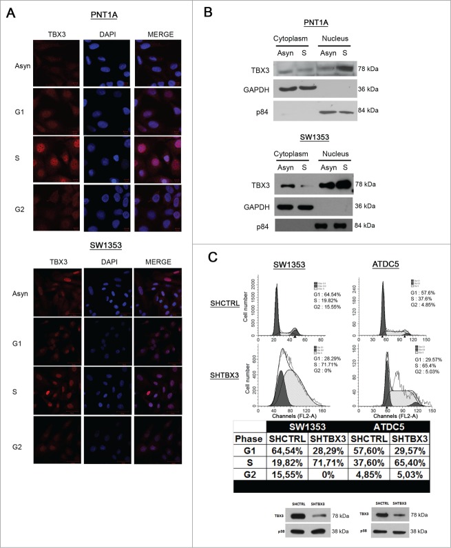
Figure 2.
TBX3 protein levels and nuclear localization are highest in S-phase and it is required for progression through S-phase into G2 (A) Immunofluorescence at 40X magnification of PNT1A and SW1353 cells using a rabbit polyclonal anti-TBX3 antibody. All cells were stained with DAPI, to determine the location of the nuclei. (B) Subcellular fractionation was performed using PNT1A and SW1353 cell lysates. Nuclear and cytoplasmic extracts were subjected to western blot analyses and probed for TBX3 using anti-TBX3 antibody. GAPDH (cytoplasmic protein) and p84 (nuclear protein) expression were determined by anti-GAPDH and -p84 antibodies. (C) Upper panel: Flow cytometry of SW1353 and ATDC5 shTBX3 and shCtrl cells. Middle panel: Table showing percentages of cells in each phase of the cell cycle. Lower panel: Knockdown of TBX3 protein in SW1353 and ATDC5 cells was confirmed by western blotting using an antibody to TBX3. Anti-p38 antibody was used as a loading control.

