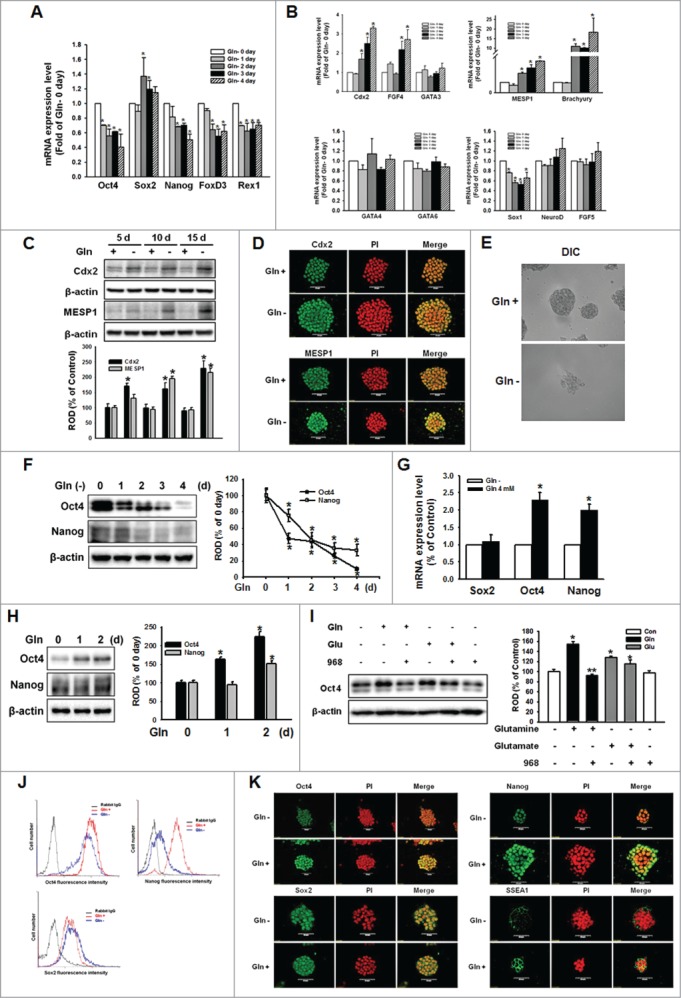
Figure 1 (See previous page).

Glutamine (Gln) deprivation deteriorated self-renewal maintenance of mESCs. The mESCs were depleted of Gln for 4 d and the total RNA was extracted as described in Materials and Methods. (A, B) The mRNA of the self-renewal marker genes (Oct4, Sox2, Nanog, FOXD3, and Rex1), trophectoderm marker genes (Cdx2, FGF4, and GATA3), endoderm marker genes (GATA4 and GATA6), mesoderm marker genes (brachyury and MESP1), and ectoderm marker genes (Sox1, NeuroD, and FGF5) were detected. Error bars represent the mean ± SEM. *P < 0.05 vs. control (0 day of Gln deprivation). (C) mESCs were cultured with/without Gln up to 15 d. The cells were passaged every 5 d. After harvest the cells, the trophectoderm marker (Cdx2) and mesoderm marker (MESP1) proteins expression was detected by Western Blot analysis. The below panel depicted by bars denote mean ± SEM of 3 experiments for each condition determined by densitometry relative to β–actin. *P < 0.05 vs. control of each day. The cells were cultured with/without for 10 d. (D) The cells were immunostained with Cdx2 or MESP1. (E) The morphological examination was performed and DIC (differential interference contrast) were acquired at 400 × magnification. (F) Total lysates subjected to SDS-PAGE, and detected Oct4 and Nanog expression. The right panel of Fig. 1F depicted by bars denote mean ± SEM of 3 experiments for each condition determined by densitometry relative to β–actin. *P < 0.05 vs. 0 day of Gln deprivation. The cells were cultured with/without Gln (4 mM) for 2 d, and (G) Sox, Oct4, and Nanog mRNA expression level were detected using real-time PCR. Error bars of (G) represent the mean ± SEM. *P < 0.05 vs. control (0 day of Gln deprivation). (H) The Oct4 and Nanog proteins expression was detected by Western Blot analysis as described in Materials and Methods. (I) The cells were pretreated with compound 968 (glutaminase inhibitor; 1 μM) for 2 hours prior to incubation with Gln (4 mM) or glutamate (Glu; 4 mM) for 2 d. And Oct4 protein expression level was detected. The right panels of (H and I) depicted by bars denote mean ± SEM of 3 experiments for each condition determined by densitometry relative to β–actin. *P<0.05 vs. Gln 0 day or control, respectively. **P < 0.05 vs. Gln alone. (J) The cells were cultured with/without Gln for 2 d and then trypsinized and suspended in PBS. After labeling with anti-rabbit Oct4 IgG, anti-rabbit Sox2 IgG, or anti-rabbit Nanog IgG, the cells were incubated with secondary conjugated antibodies (goat-anti-rabbit IgG-conjugated FITC) and detected with flowcytometry. (K) The cells were double labeled with anti-Oct4, -Nanog, Sox2, or -SSEA1, and counter stained with PI. And then the cells were observed with a confocal microscope.
