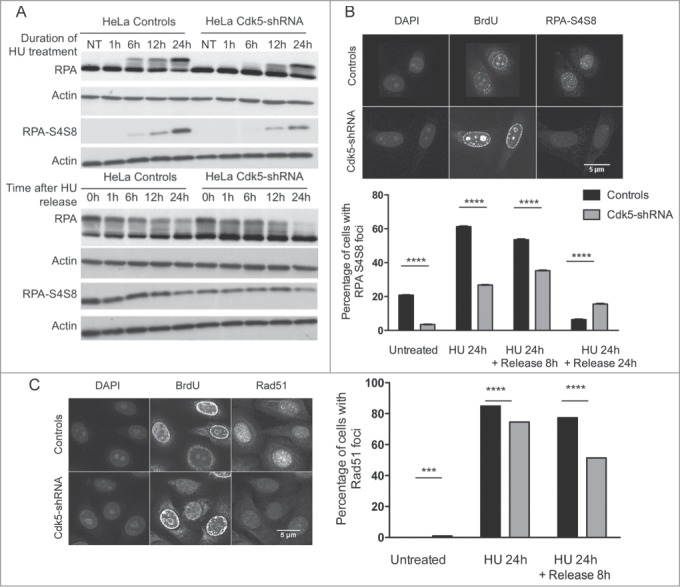
Figure 5.

Lower RPA S4S8 phosphorylation and Rad51 foci formation in Cdk5-shRNA cells after HU treatment. (A) Representative western blot of phospho-RPA S4S8 levels in protein extracts from Control and Cdk5-shRNA during HU block (2 mM,) and up to its removal from the culture medium. RPA S4S8 was detected using a phosphospecific antibody. Actin expression was used as a gel loading control. (B) Phospho-RPA foci formation: Cells were released from HU (2 mM, 24 h) at the indicated times, fixed and immunostained for RPA S4S8. The figures are representative of 2 independent experiments with 2 different clones. The mean percentage of cells carrying more than 5 foci are presented, with SEM. Only BrdU positive cells (S-phase) were quantified with 350 to 600 cells analyzed (****P < 0.0001; P < 0.0001, unpaired t-test). (C) Formation and persistence of RAD51 foci in Cdk5-shRNA and Control cells. Cells were treated or not with HU (2 mM, 24 h) and Rad51 foci quantified using a 2D Spinning-Disc/Tirf/Frap system after a pulse labeling with BrdU (10 μM, 15 min) at the indicated times. Data are representative of 4 independent experiments using 2 different Cdk5-shRNA clones with between 600 and 3,000 cells analyzed for each condition and represent the number of BrdU positive cells carrying more than 5 foci, compared by t-test between the different treatment groups (***P < 0.001; ****P < 0.0001).
