Figure 1.
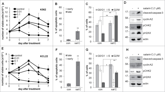
Salarin C inhibits cell proliferation and induces apoptosis and DNA damage in CML cell lines. K562 (A) or KCL22 (E) cells were plated at 3×10⁵ cells/ml and after 24 hours (time 0) were treated or not (control) with a single dose of salarin C at the indicated final concentration (µM); cells were then incubated in normoxia and trypan blue-negative cells were counted at the indicated timepoints; values are averages ± SEM of data from 3 independent experiments; significant differences are indicated (Student's t test for independent samples; *: p< 0.05. K562 (B, C) or KCL22 (F, G) cells were incubated as above in the presence (sal C) or not (con) of 1 µM salarin C and subjected to Annexin V / propidium iodide assay to determine the percentages of cells in early or late apoptosis (B, F) or labeled with propidium iodide alone (C, G) to determine cell cycle phase distribution. Analysis was performed by flow cytometry at day 3 of incubation; values are averages ± SEM of data from 3 independent experiments; significant differences are indicated (Student's t test for independent samples; *: p< 0.05, **: p< 0.01). K562 (D) or KCL22 (H) cells were lysed at day 3 of incubation and lysates subjected to immuno-blotting with antibodies raised against the indicated proteins; anti-actin antibody was used to verify equalization of protein loading; one representative experiment out of 3 is shown.

