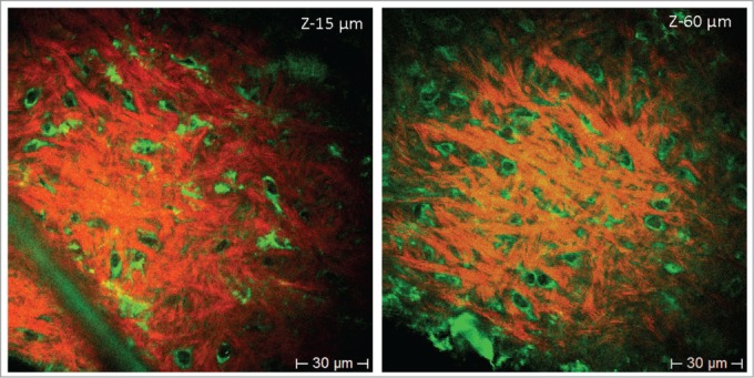
Figure 3.

Label-free HAP stem cells in the bulge of the mouse whisker imaged with multiphoton tomography. Images are produced due to the 2-photon-excitation-induced autofluorescence of cells and SHG of collagen fibrillar structures at different depths (z-15 µm and z-60 µm, 760 nm excitation). In these autofluorescence images, fluorescence is detected mainly in the cytoplasm including extrusions, but not in the nucleus. Label-free HAP stem cells within the hair follicle bulge have the same morphology and size as the ND-GFP HAP stem cells with a round-shaped body and with typically 2–3 extrusions (green color). Collagen is imaged by SHG (red [pseudo color coded]).
