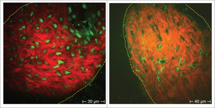
Figure 4.

ND-GFP-expressing (left) and label-free HAP stem cells (non-GFP, right) in the dermal papilla. HAP stem cells were imaged with 2-photon induced GFP or by autofluorescence, respectively. Extracellular matrix protein collagen was imaged due to SHG. In the GFP fluorescence image the entire HAP stem cell fluoresced bright green (left panel). In the autofluorescence image (right panel), the cell nucleus appeared dark due to the lack of endogenous fluorophores. Bright autofluorescent structures are in the mitochondria within the cytoplasm, which are rich in intrinsic fluorescent NAD(P)H and flavins. Collagen fibers visualized by SHG are well aligned structures (red [pseudo color coded]).
