Figure 2.

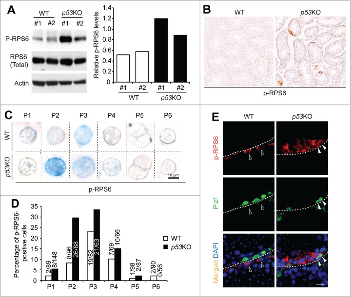
p53 loss amplifies mTORC1 activity in differentiating spermatogonia. (A) western blotting analysis of p-RPS6 from 2 mice per genotype (#1 and #2) (left). Quantification of the relative levels of p-RPS6 against total RPS6 (right). (B) immunohistochemistry staining of p-RPS6 in 4-weeks old juvenile WT and p53KO testis. Images shown are representative from 3 mice per genotype examined. (C) immunohistochemistry staining of p-RPS6 detected by alkaline phosphatase (AP) in cells from P1 to P6 populations isolated from 4-weeks old juvenile WT and p53KO pStra8-GFP mice. Cells shown are magnified views of a single cell. Number of positive cells detected and total number of cells examined are indicated. (D) comparison of the percentages of p-RPS6-positive cells in P1 to P6 populations. Graphs represent mean values ± SEM. n = 3 mice per genotype, *P < 0.05. (E) dual-immunofluorescence staining of p-RPS6 (red) and Plzf (green) in 4-weeks old juvenile WT and p53KO testis.
