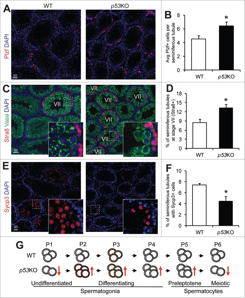
Figure 4.

p53 loss expands the spermatogonia pool but causes a loss of meiotic spermatocytes. A) immunofluorescence staining of Plzf in 4-weeks old juvenile WT and p53KO testis. (B) quantification of cells showing strong positivity for Plzf in seminiferous tubules of testis from (A) representing early differentiating spermatogonia. Graphs represent mean values ± SEM. n = 4 mice per genotype, *P < 0.05. (C) dual-immunofluorescence staining of Stra8 (red) and Vasa (green) in 4-weeks old juvenile WT and p53KO testis. (D) quantification of percentage of stage VII seminiferous tubules in cross sections as shown in (C). Graphs represent mean values ± SEM. n = 4 mice per genotype, *P < 0.05. (E) immunofluorescence staining of Sycp3 in 4-weeks old juvenile WT and p53KO testis. (F) quantification of the percentage of seminiferous tubules containing meiotic spermatocytes showing strong positivity for Sycp3 from panel E. Graphs represent mean values ± SEM. n = 4 mice per genotype, *P < 0.05. (G) schematic diagram showing the effect of p53 deletion on spermatogonial differentiation. Orange and red colors in cells at P2 and P3 populations indicate mTORC1 activity.
