Figure 3.
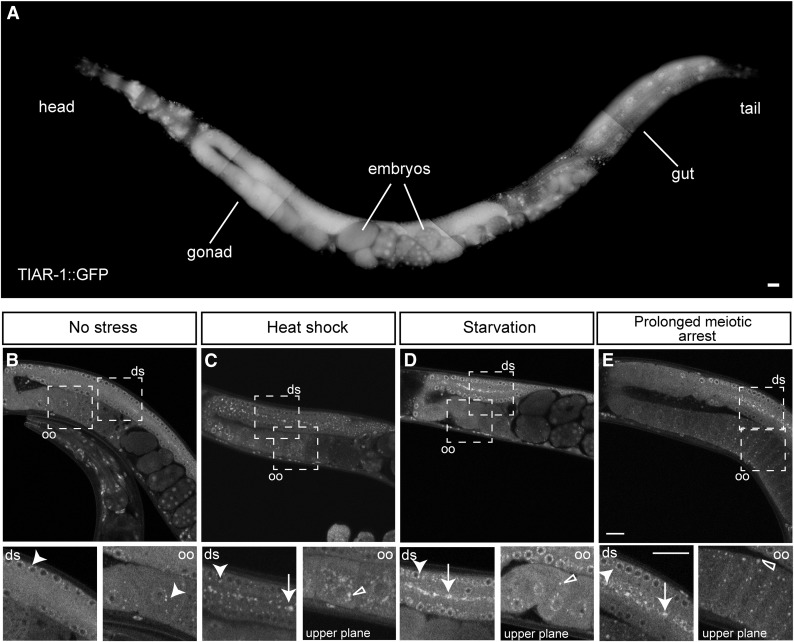
TIAR-1 associates with cytoplasmic granules during stress in the gonad. TIAR-1::GFP expression and localization were assessed in tiar-1(tn1545). (A) 1-d-old tiar-1::gfp hermaphrodites were anesthetized and observed with the fluorescence microscope. The subcellular localization of TIAR-1::GFP in the gonad was observed under normal growth conditions (B), after exposure to heat shock (3 hr at 31°) (C), and after starvation (4 hr) (D). TIAR-1::GFP subcellular localization was also observed in tiar-1(tn1545); fog-2(q71) unmated females (E). These animals were imaged using confocal microscopy. Dotted squares indicate the zoomed-in areas. ds, distal gonad; oo, oocytes. Arrowheads indicate likely P granules, arrows gonad core granules, and empty triangles oocytes granules. Scale bars, 10 µm.

