Figure 9.
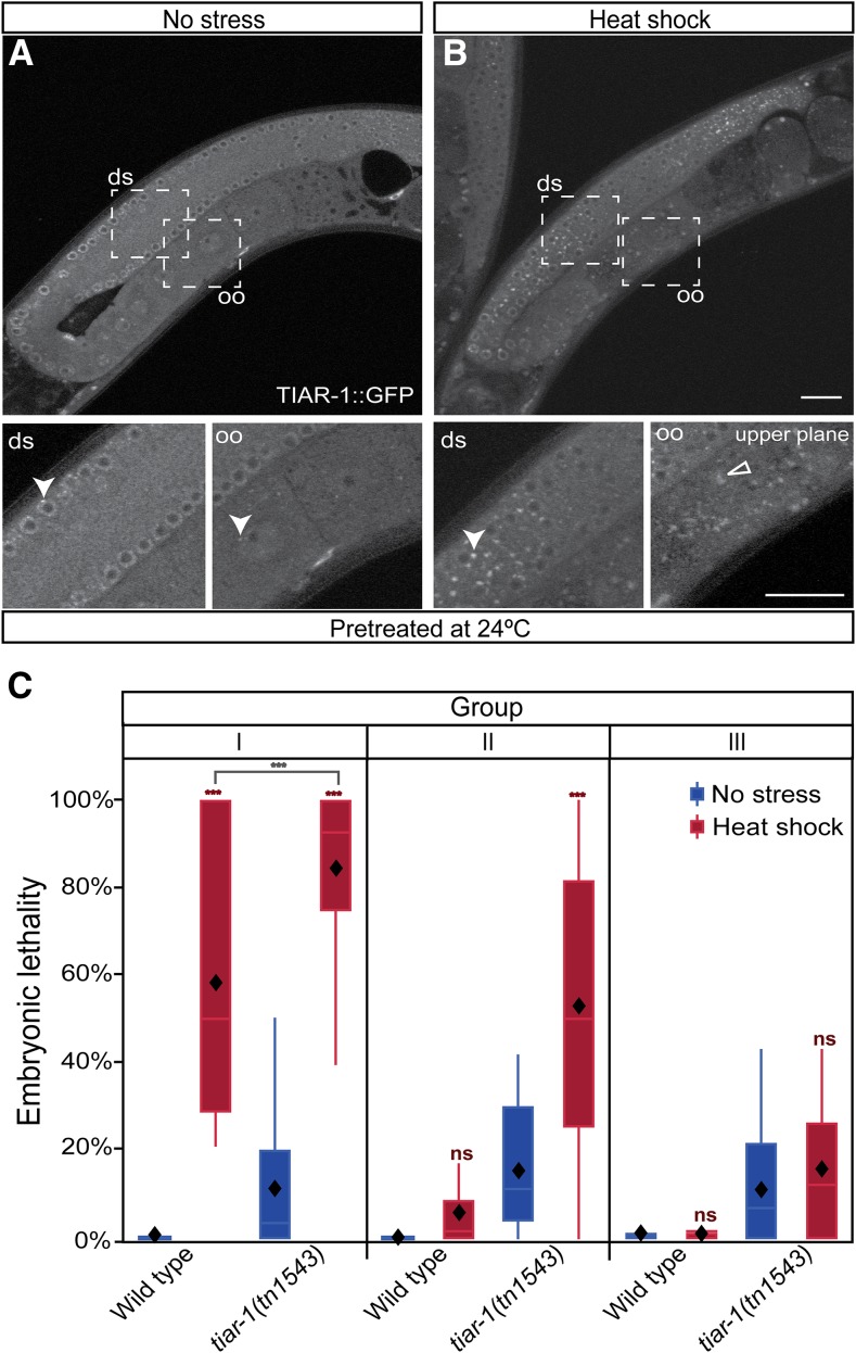
tiar-1 protects female germ cells and embryos from heat shock independently of gonad core granules. Pretreated tiar-1::gfp animals (grown at 24°) were imaged using confocal microscopy. Representative images show gonads of animals (A) not exposed to stress, and (B) exposed to heat shock (31° for 3 hr). The photomicrograph in (B) is representative of animals that formed very small granules in the core gonad and normal granules in oocytes. However, most of the pretreated animals did not form core granules after the heat shock (Table 4). Arrowheads indicate likely P granules and empty triangles oocytes granules. Scale bar, 10 µm. (C) Pretreated wild-type and tiar-1(tn1543) hermaphrodites were exposed to heat shock (3 hr at 31°) or kept at 20° as a control. After, their progeny were sorted into three groups (as in Figure 7A). Embryonic lethality was quantified for all the groups (C). Embryos not hatching within 24 hr after being laid were considered inviable. The graphs show the data obtained from three independent replicates. The tiar-1(tn1543) animals included in this experiment were representative of the total population (see Table 4); we did not prescreen or exclude the small fraction of animals containing small granules in the gonad core. The boxes represent the 25–75% interquartile range, the diamonds the mean value, and the whiskers extend from the upper to the lower values. The *** (P < 0.001) and ns (nonsignificant) symbols placed on top of the boxes are comparisons between no-stress and stress groups, whereas the ones placed above them are comparisons between the stress groups. Least square means, Tukey HSD test.

