Figure 1.

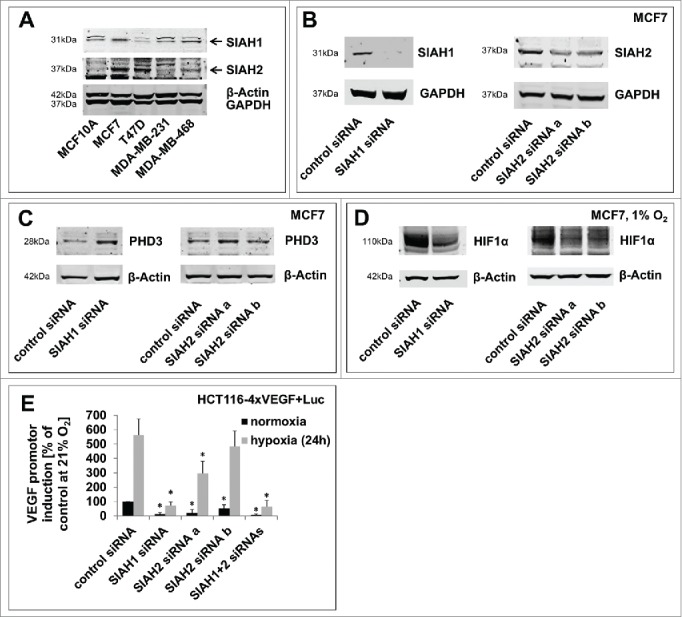
SIAH1/2 silencing reduces hypoxic adaptation in breast cancer cells. (A) Comparison of SIAH1 and SIAH2 expression levels in different breast cancer cell lines. Four breast cancer cell lines and MCF10A as a non-cancer control cell line were lysed and immunoblotted for SIAH1 expression. The membrane was reprobed for SIAH2. GAPDH and β-Actin serve as a loading control. (B) Efficient SIAH knockdown in MCF7 cells. MCF7 breast cancer cells were transfected with siRNAs targeting SIAH1 or SIAH2. After 48 h the cells were lysed and Western Blot was performed with specific antibodies. Instead of reprobing the membrane was cut in advance to allow clear visualization of the results. (C) Lysates of MCF7 cells silenced for expression of SIAH1 or SIAH2 were examined for expression of PHD3. (D) MCF7 cells were transfected with SIAH1 and SIAH2 siRNA and cultivated at 1% O2 for 48 h. Lysates were probed for HIF1α accumulation. (E) SIAH1 and SIAH2 were silenced in HCT116–4xVEGF+Luc cells for 24 h. The cells were then incubated at 21% O2 or 1% O2 for another 24 h, lysed, and a luciferase assay was performed to assess HIF target gene expression. Relative light units from the luciferase assay were normalized to the cell number measured with CellTiter-Blue Cell Viability assay. Bar graphs show mean values, error bars indicate SD. Data were analyzed using 1-way ANOVA followed by Dunnett's post-hoc test. n = 3; *, p<0.05.
