Figure 2.

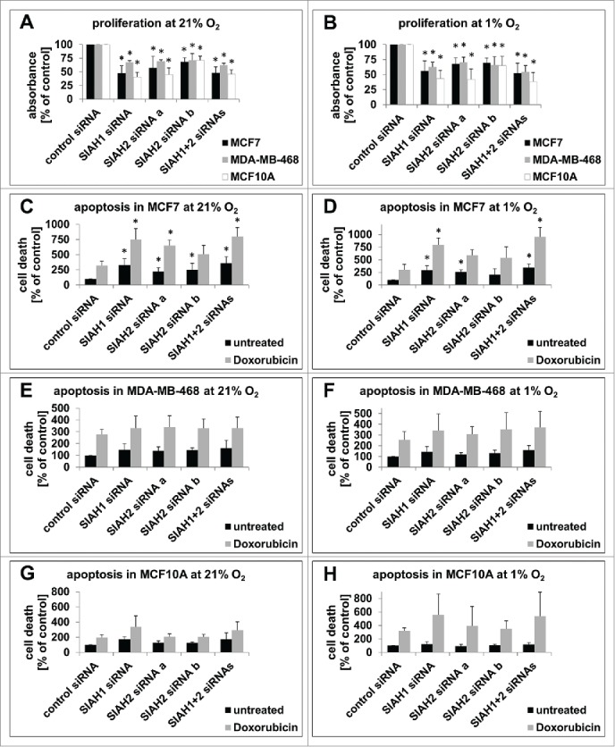
SIAH1/2 silencing inhibits proliferation and promotes apoptosis in breast cancer cells. (A, B) 48 h after transfection of MCF7, MDA-MB-468, and MCF10A cells with RNAi targeting SIAH1/2, cells were incubated in presence of BrdU for 2 h. BrdU incorporation into the DNA, signifying cell division, was quantified with an ELISA. Cells were either kept at 21% O2 (A) or 1% O2 (B) for the duration of the experiment. (C, D) 30 h post RNAi transfection, MCF7 cells were either left untreated or apoptosis was stimulated by adding the DNA-damage inducing chemotherapeutic drug Doxorubicin (1 µM) for 18 h. Then cells were lysed and apoptosis was measured with a TUNEL-based ELISA. The same experiments were performed with MDA-MB-468 (E, F) and MCF10A cells (G, H). Cells were either kept at 21% O2 (C, E, G) or 1% O2 (D, F, H) the whole time. Bar graphs show mean values, error bars indicate SD. Data were analyzed using 1-way ANOVA followed by Dunnett's post-hoc test. n = 3; *, p<0.05.
