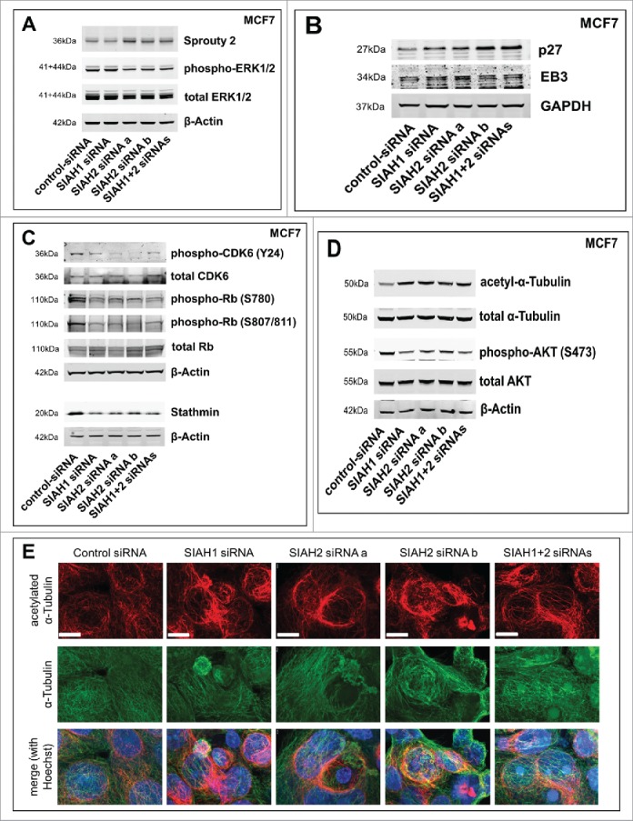
Figure 4.

SIAH1/2 silencing affects expression of p27 and stathmin in breast cancer cells. (A) Following siRNA transfection (48 h), MCF7 cells were lysed. Western blot was performed to assess expression levels of the SIAH substrate Sprouty2 and activation status of ERK1/2. (B) Western Blot showing expression levels of the SIAH substrates p27Kip1 and EB3 after silencing of SIAH1 or SIAH2. (C) Western blot demonstrating the effect of SIAH1/2 silencing on total CDK6 levels and CDK6 phosphorylation at tyrosine 24, total Rb levels and Rb phosphorylation at serines 780 and 807/811, as well as on stathmin protein levels. (D) Western blot to determine the stability of microtubules (via acetylated α-Tubulin) as well as the activity of AKT (phosphorylation at serine 473) downstream of stathmin. (E) Immunofluorescent staining to determine microtubule stability after knockdown of SIAH1 and SIAH2. Following RNAi transfection (48 h), MCF7 cells were fixed and stained for α-Tubulin (DyLight 488, green) and acetylated α-Tubulin (Cy3, red). Cell nuclei were stained with Hoechst dye (blue). Scale bars, 10 µm.
