Figure 6.

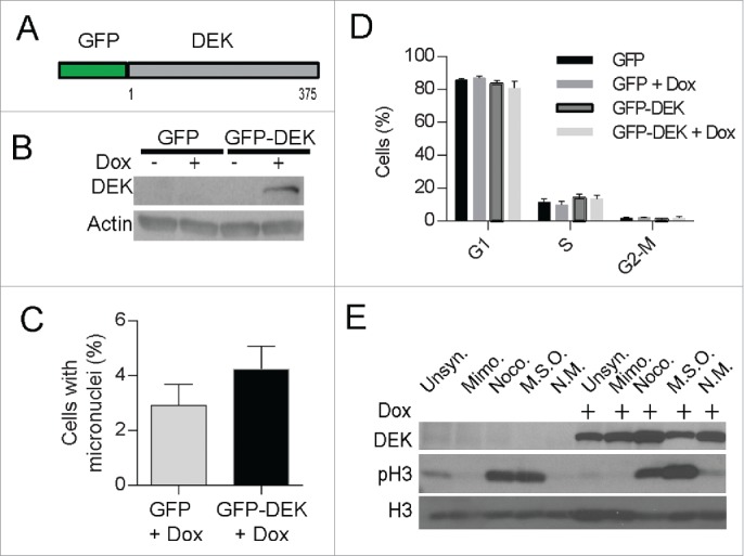
Acute, inducible expression of DEK induces micronuclei formation. Dek−/− MEFs were transduced with a doxycycline-inducible GFP-DEK fusion protein (A) encoded by a TRIPZ vector and selected with puromycin. The empty GFP pTRIPZ vector was used as a control. (B) Western blot analysis confirmed fusion protein expression at 48 hours after the addition of doxycycline to GFP-DEK transduced cells. Cells from (B) were fixed on coverslips and micronuclei were quantified by IF as before (C). (D) GFP-DEK expression does not alter cell cycle progression. GFP and GFP-DEK transduced cells, in the presence or absence of dox for 48 hours, were pulsed with EdU for 2 hours and subjected to cell cycle analysis using flow cytometry. Cells in each stage of the cell cycle were quantified from duplicate experiments. (E) The GFP-DEK fusion protein is retained in mitotic cells. Western blot analysis of GFP-DEK cells with and without doxycycline and arrested in G1 with mimosine or mitosis with nocodazole, or underwent a mitotic shake off (MSO) confirms an increase of GFP-DEK in mitotic cells (NM=non mitotic). A DEK specific antibody was utilized (BD Biosystems) as well as a pH3 antibody to detect cells in mitosis.
