Figure 7.

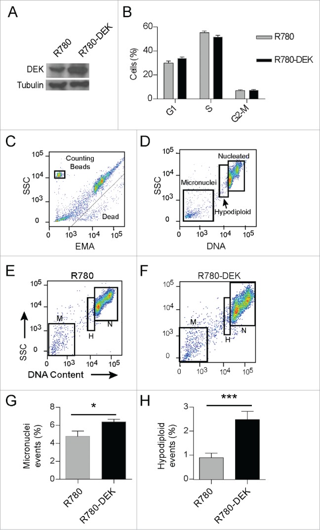
DEK over-expression increases the incidence of hypodiploidy and micronuclei in cancer cells. CCHMC-SCC1 cells are a squamous cell carcinoma line isolated from a primary tonsillar tumor. (A) DEK was over-expressed by R780-DEK compared to control R780 transduction, and expression levels were confirmed by protein gel blot analysis. (B) DEK overexpression did not increase proliferation in C-SCC1 cells. Cells were pulsed with EdU for 2 hours, and then stained with 7AAD to determine DNA content. (C-F) CCHMC-SCC1 cells treated as in (A) were used for micronuclei detection by a flow cytometry based assay. (C) DNA from dead or dying cells was gated out on EMA positivity. (D) Remaining DNA that was stained with SYTOX Red was gated on DNA content to reveal G1, G2/M nuclei (labeled nucleated or N), a hypo-diploid population of nuclei (labeled H), and micronuclei (M). ((E)and F) Each population of events was gated in FlowJo to compare empty vector control cells (E) with DEK overexpressing cells (F). (G and H) DEK over-expression increased both micronucleus formation (G) and the hypo-diploid population of cells (H). Numbers represent 5 replicate cell cultures from 2 independent experiments.
