
Figure 2.
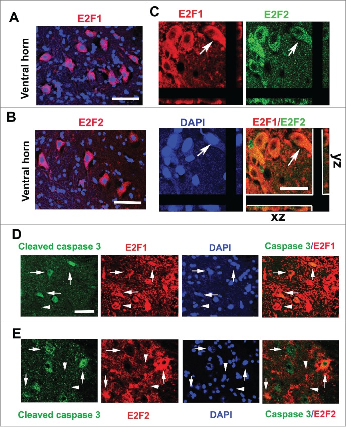
Upregulated E2F1 and E2F2 are associated with cleaved caspase-3+ neurons at 24 hours after SCI. (A-B) E2F1 and E2F2 are induced in neurons of the ventral horn (VH) gray matter at day 1 after SCI. (C) Most of E2F1+ cells (red) in the gray matter were also co-labeled with E2F2 (green), which had neuronal morphology. Arrows indicated E2F1+/E2F2+ cells with z-stacks (x-z axis, y-z axis). (D-E) Co-localization between cleaved caspase-3 (green) and E2F1 (red) or E2F2 (red) is apparent in the VH at day 1 after SCI. Arrows indicated caspase-3+/E2F1-2+; Arrowheads indicated caspase-3−/E2F1-2+. Scale bar is 100 μm.