Figure 3.
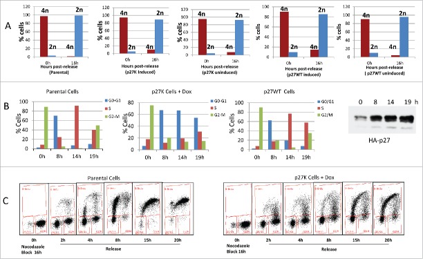
U2OS cells, U2OS-p27WT cells and U2OS-p27K cells were placed in nocodazole inhibition as described in Material and Methods. Some cultures of U2OS-p27K and U2OS-p27WT cells had Dox (1µg/ml) added for 6 h. Cultures of parental, non-induced U2OS-p27WT, non-induced U2OS-p27K, and induced U2OS-p27K cells were released from the nocodazole block. Panel A shows propidium iodide straining of these cells, as described in Material and Methods, before release and 15 h after release from the mitotic inhibition. Data show 2N and 4N staining. Panel B shows the cell cycle distribution with propidium iodide staining of parental cells, U2OS-p27WT, and U2OS-p27K induced 6 h before mitotic release. Panel C illustrates the induction of p27K by 1 µg/ml of Dox. Panel D shows the analysis of BrdU incorporation in cells treated as in Panel B when released into fresh medium containing 30 μM BrdU.

