
Figure 4.
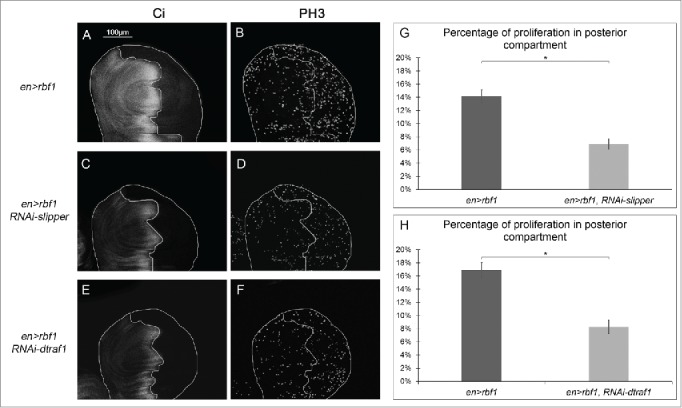
Cell proliferation induced in response to Rbf1-apoptosis requires Slipper and dTRAF1 (A, C, E) anti-Ci staining used to visualize the anterior domain in wing pouch imaginal discs from en-Gal4/+; UAS-rbf1/+ or en-Gal4/UAS-RNAi slipper ; UAS-rbf1/+ or en-Gal4/+; UAS-rbf1/UAS-RNAi dtraf1 third instar larvae. A line indicates the antero-posterior frontier and the posterior compartment is on the right side. (B, D, F) Visualization of mitotic cells by PH3 staining in wing pouch imaginal discs from the previously described genotypes. (G, H) Comparison of proliferation percentage in posterior compartment between wing pouch imaginal discs from en-Gal4/+; UAS-rbf1/+ and en-Gal4/ UAS-RNAi slipper ; UAS-rbf1/+ third instar larvae or between en-Gal4/+; UAS-rbf1/+ and en-Gal4/+; UAS-rbf1/UAS-RNAi dtraf1 third instar larvae. Asterisks indicate a statistically significant difference between 2 genotypes (Student's t-test α < 0.05). Each experiment presented in B, and H was independently performed 3 times.