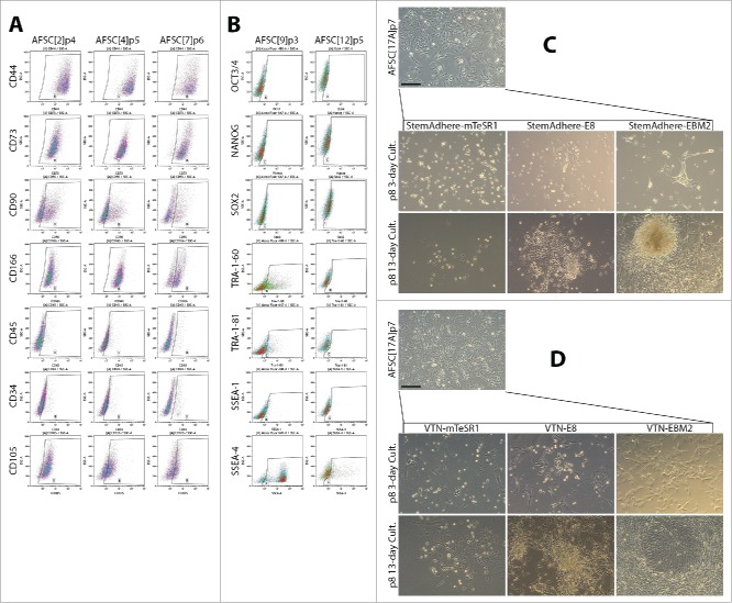
Figure 1.
Characterization of AFSC and optimization of the culture condition following episomal reprogramming. (A) Expression of MSC markers in AFSC measured by flow cytometry. The expression pattern is similar to a typical MSC pattern, with some variability in CD90 and CD166. CD34 and CD45 expression were low. (B) Flow cytometric analysis of ESC marker expression in AFSC. No Oct3/4 expression was observed, Nanog expression was negligible, dim Sox2 signal was seen in over 30% of the cells. TRA antigen expression was observed in AFSC9 but not AFSC12. SSEA-1 expression was negligible, a bright SSEA-4 signal was found to be high in AFSC9 but much lower in AFSC12. The pattern of ESC marker expression is not consistent with pluripotency, (C, D). Morphological transformation of AFSC in response to episomal plasmid reprogramming, secondary passaging on day 9 and subsequent culturing on StemAdhere-coated plates in 2 different pluripotency-supporting media – mTeSR-1, E8. AFSC growth medium was used as a control (C); and on VTN-coated plates in the same media (D). VTN in combination with the E8 medium supported emerging colonies of epithelioid cells while StemAdhere and mTeSR-1 medium were non-permissive. Scale bars = 200 µm.

