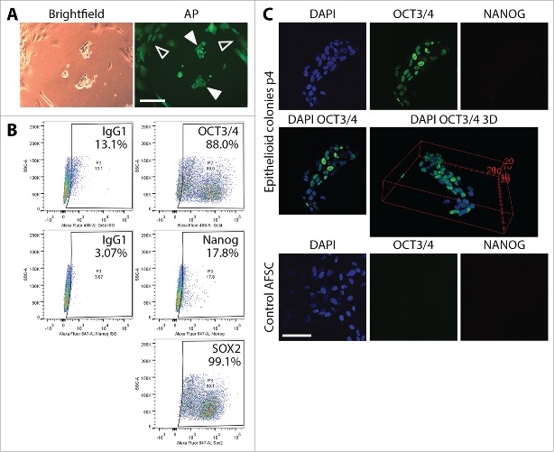
Figure 2.
Characterization of partially reprogrammed epithelioid cells resulting from episomal reprogramming of AFSC. (A) Alkaline phosphatase staining of colonies of partially reprogrammed cells before mechanical picking and expansion. Discreetly bright AP signal beyond background fluorescence (hollow arrows) was not observed, confirming incomplete reprogramming (filled arrows). (B) Flow cytometric analysis of Oct4, Nanog and Sox2 in mechanically picked epithelioid colonies cultured for 14 days (30 days post-transfection) on VTN-coated plates in E8 medium. The profile corroborated incomplete reprogramming. Scale bars = 200µm. (C) Immunocytochemical staining and confocal microscopy visualization of ESC markers Oct4 and Nanog in epithelioid cell colonies expanded for 4 passages post-transfection. Oct4 expression was clearly heterogeneous among the individual cells in the colonies ranging from cells where Oct4 is completely absent to Oct4 being represented by a strong fluorescent signal. Nanog expression was not observed. As a control, we stained unreprogrammed AFSC for the same markers. These cells exhibited no Oct4 and Nanog expression at all. Scale bar = 50 µm.

