
Figure 3.
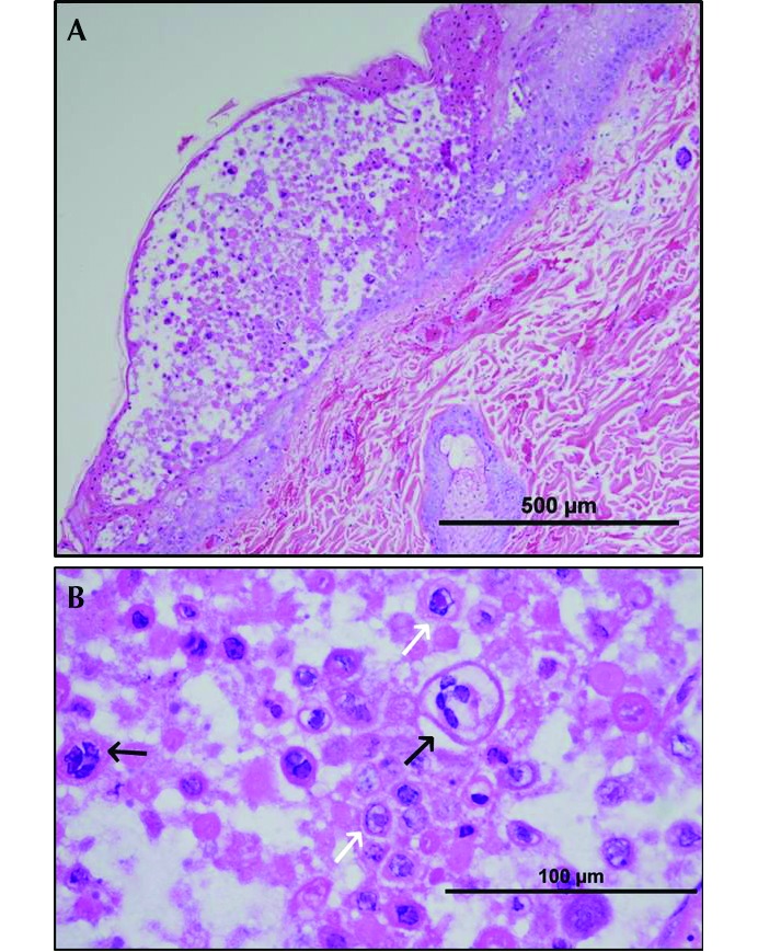
(A) Haired skin: degeneration and necrosis with vesicle formation. (B) Haired skin (higher magnification): necrotic epithelial cells, epithelial syncytia (black arrows), and eosinophilic intranuclear inclusions (white arrows). Hematoxylin and eosin stain.